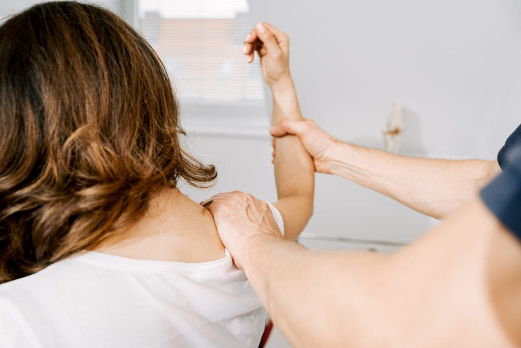
Orthopaedics treats chronic illnesses and acute injuries to the musculoskeletal system. The following components are important as part of our multimodal complex therapy:
In order to be able to use these therapy methods effectively from the outset, 4D spinal column measurement, movement analysis and biofeedback are used so that incorrect static posture and/or muscular imbalances can be eliminated – with the help of orthopaedic aids if necessary. In addition, psychological, social and occupational aspects must always be taken into account in the treatment concept. The use of psychotherapeutic measures (talk therapy, relaxation techniques) is of enormous therapeutic benefit, especially in the case of chronic pain.
Injection therapies are among the fastest and most effective methods of orthopaedic pain therapy. By injecting pain-relieving, anti-inflammatory and anti-inflammatory drugs – including biological and naturopathic remedies – directly into the source of the pain, it is possible to influence the underlying disorder without burdening the body with more medication than necessary.
A distinction is made between subcutaneous, intramuscular and intraarticular injections as well as injections into bursae or tendon insertions.
Autologous blood therapy is one of the treatment methods used in alternative medicine. In this procedure, the patient’s own blood is taken, prepared in various ways and then re-injected. This procedure is effective for a large number of overuse injuries, injuries, wear and tear and to accelerate healing after operations.
A distinction is made between the body’s own blood components ACP (Autologous Conditioned Plasma) and PRP (Platelet Rich Plasma). The basis for the success of the ACP or PRP treatment used is the knowledge that wound healing factors are present in blood plasma and blood platelets (thrombocytes) in a certain concentration.
Specifically, these wound healing factors are growth factors that lead to improved cell regeneration, increased collagen production and improved blood supply to the tissue. On the other hand, they are anti-inflammatory factors that contribute to the reduction of tissue inflammation in wear processes and injuries.
The still relatively young field of regenerative medicine deals with the healing of various diseases by restoring dysfunctional cells, tissues and organs both through biological replacement – for example with the help of cultivated tissue – and by stimulating the body’s own regeneration and repair processes through growth factors.
In orthopaedics, bioregenerative therapy is used to harvest stem cells from fatty tissue. With the help of this minimally invasive procedure, the use of an artificial joint can often be delayed – or even avoided completely. This method is currently only approved for use on the knee joint.
Shock wave therapy is a newer form of treatment that promises gentle treatment of inflammation, calcifications or injuries in many cases.
Shockwave therapy also has virtually no side effects and makes many operations unnecessary.
Shock waves are short, high-energy mechanical-acoustic waves that are transmitted through water or water-containing tissue without attenuation. Elastic body tissue – including muscles and fatty tissue – also simply transmits the shock waves. Only when the shock wave hits solid tissue components – kidney stones, gallstones, calcium deposits in tendons – is the energy contained in the shock wave discharged. It acts on the solid resistance in the tissue and leads to the mechanical disintegration of foreign bodies. Orthopaedics in particular has developed and tested a wide range of applications for shock wave treatments.
These include:
Shock wave therapy can not only shatter calcifications, but also treat tendon attachment inflammation or heal poorly healing bone fractures. Shock waves generally accelerate the healing process in tissues because they also promote the formation of the body’s own messenger substances, which promote healing processes.
Effects of shock wave therapy
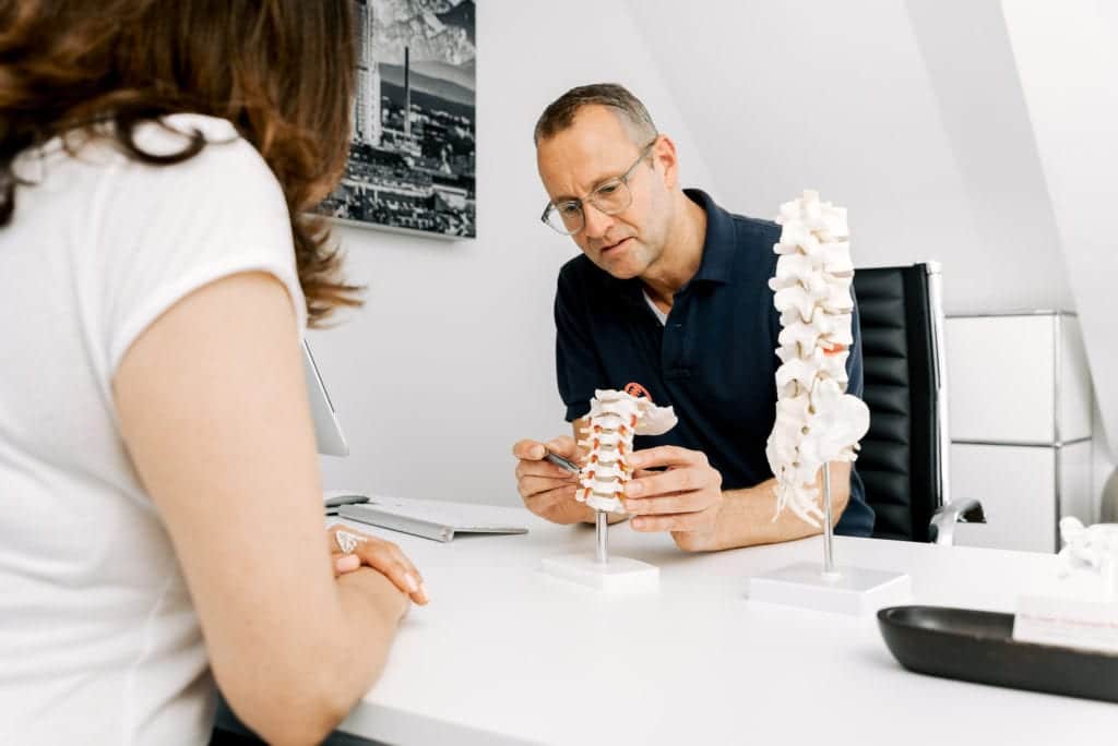
The causes of back pain can be very diverse and therefore require an interdisciplinary treatment concept. The first step in making a diagnosis is to question the patient in detail and carry out an intensive physical examination. Other special diagnostic procedures such as ultrasound, magnetic resonance imaging (MRI), computer tomography (CT) and scintigraphy are essential. As there is no direct correlation between the degree of symptoms and the radiological findings, a neurological examination with electromyography (EMG) and measurement of nerve conduction velocity (NLG) is often useful and necessary for verification.
Only after an exact diagnosis has been made is an individual treatment plan drawn up for the patient, during the course of which pain and its causes are eliminated. Minimally invasive treatment methods such as image-guided infiltrations, epidural catheters, radiofrequency therapy and microlasers are used alone or in combination. As a rule, these can be carried out on an outpatient basis. These procedures are supplemented by physiotherapy and medication.
This outpatient procedure is used for complaints caused by osteoarthritis of the small intervertebral joints in the entire spine and as a diagnostic aid prior to radiofrequency therapy (see below). Typical complaints are morning stiffness or back pain after getting up in the morning or from a sitting position. The pain is often described as “belt-shaped”.
A special needle is used to apply anti-inflammatory and pain-relieving medication to the affected joints under image converter or CT guidance. This injection can also be repeated several times as required.
The aim is to achieve the maximum effect with a minimal outpatient procedure. These procedures are also used to treat irritation of the nerve roots in the cervical, thoracic and lumbar spine caused by bulging or herniated discs, narrowing of the vertebral and/or nerve canal, as well as complaints following disc surgery (“post-nucleotomy syndrome”) or fusion surgery.
Under image converter or CT control and, if necessary, with contrast medium, the irritated root and/or the natural opening at the transition from the coccyx to the sacrum is precisely infiltrated into the spinal canal. Precise alignment of the needle tip using image-guided therapy is an important prerequisite for the effectiveness of the therapy applied. It avoids inaccurate placement of the drug depot in
unrecognized soft tissue and at the same time reduces the required drug dose to the necessary minimum. Successful treatment interrupts the pain spiral through a local drug effect and ensures rapid and lasting success through a decongestant and anti-inflammatory effect. The infiltration should be carried out several times at intervals of several days or weeks.
The advantage of these procedures is the rapid mobilization with the possibility of starting light physical work as well as the repeatability or combination of therapy modules.
This procedure is a low-risk alternative to open disc or spinal surgery and is used for bulging or herniated discs, post-nucleotomy syndrome or narrowing of the spinal or nerve canal in the cervical, thoracic or lumbar spine, in each case with acute or chronic pain radiating into the arms, buttocks or legs.
It is performed on an outpatient basis or as part of a short inpatient stay of two to three days in a “twilight sleep”. A thin special catheter with a spiral spring at the end is inserted into the spinal canal via a guide needle through the natural opening in the area of the coccyx. This allows the catheter to be advanced without injury. Here too, the catheter tip is placed exactly at the site of the pain under image converter control. The procedure takes 30 to 45 minutes. Saline, enzymes and pain-relieving and anti-inflammatory medication are administered via the catheter.
The aim of the treatment is to shrink bulging intervertebral disc tissue, alleviate pain, eliminate inflammation of the tissue and loosen inflammation-related adhesions of the various structures. The catheter remains in place for at least 48 hours and is not perceived by the patient as painful or disruptive to movement. Injection of the drug combination is generally followed by a short period of rest at the patient’s bedside, after which the patient is advised to rest for one to two weeks. In most cases, light work can be resumed immediately after consultation with the attending physician
If the above-mentioned injection of the small vertebral joints does not lead to a permanent reduction in pain in the case of degenerative changes, the nerve transmitting the pain can be interrupted in the long term with a “heat probe”.
Radiofrequency therapy/thermocoagulation is a less invasive procedure that can be performed on an outpatient basis. After precise positioning of the specially developed needle under image converter control, the position is checked again with the help of a short stimulation current pulse. After a local anaesthetic, the tip of the probe is heated for 90 seconds to switch off the nerve and, as each individual vertebral joint is supplied by several nerve fibers, the therapy must be carried out on several levels. The treatment does not cause any impairment of physical functions.
The elimination of pain symptoms is usually permanent. If the nerve tissue recovers after one to two years, the procedure can be repeated without any problems. It is also possible to carry out light physical work without restriction.
This gentle, minimally invasive procedure can be used for protrusions, minor prolapse or wear of the disc in the cervical or lumbar spine, with or without pain. It is another alternative to conventional open disc surgery without the risk of scarring.
A very fine special probe is inserted directly into the painful intervertebral disc under constant position monitoring using an image converter or CT. A test injection using a contrast medium (“discography”) ensures that the pain also originates from this damaged disc. The laser fiber is then inserted into the intervertebral disc via the probe.
The laser energy has several effects: by partially vaporizing the gelatinous disc nucleus and the resulting shrinkage of the disc, the nerve is immediately relieved and the excruciating nerve and/or back pain is eliminated. In addition, pain-conducting fibers in the intervertebral disc tissue are obliterated and pain transmission substances to the brain are eliminated. Tears in the tissue of the intervertebral disc are also closed, so that it solidifies and stabilizes again.
This procedure is also performed on an outpatient basis under local anesthesia; if necessary, a “twilight sleep” can also be performed. The patient is then given a special corset for six weeks to relieve pressure on the spine. Light work can be resumed immediately, but physically strenuous activities and sport require rest for three to four weeks. Supportive physiotherapy should be initiated after two weeks.
You are currently viewing a placeholder content from Vimeo. To access the actual content, click the button below. Please note that doing so will share data with third-party providers.
More InformationYou are currently viewing a placeholder content from YouTube. To access the actual content, click the button below. Please note that doing so will share data with third-party providers.
More InformationYou need to load content from reCAPTCHA to submit the form. Please note that doing so will share data with third-party providers.
More InformationYou are currently viewing a placeholder content from Facebook. To access the actual content, click the button below. Please note that doing so will share data with third-party providers.
More InformationYou are currently viewing a placeholder content from Instagram. To access the actual content, click the button below. Please note that doing so will share data with third-party providers.
More InformationYou are currently viewing a placeholder content from Google Maps. To access the actual content, click the button below. Please note that doing so will share data with third-party providers.
More InformationYou are currently viewing a placeholder content from X. To access the actual content, click the button below. Please note that doing so will share data with third-party providers.
More Information